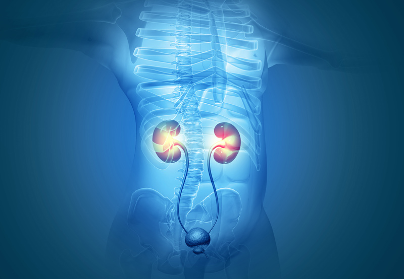

Ureteral obstruction may be caused by an internal ureteral blockage inside or outside of the ureter which can be at any level of the ureteral length. This can include ureteropelvic junction (UPJ) obstruction, or ureteral strictures at any level of the ureter from kidney junction to the bladder junction. In some cases, the obstruction is due to a mechanical effect (like a mass) adjacent to the ureter. Our experts at UCI Urology will evaluate the underlying reason and address it as needed.
Ureteropelvic Junction Obstruction
UPJ obstruction is when part of the kidney is blocked, typically at the renal pelvis. This is where the kidney attaches to the ureter, a tube that carries urine to the bladder. The blockage may slow or completely stop the flow of urine out of the kidney, leading to buildup that damages the kidney. In some cases, surgery may be necessary to address the problem.

How the Kidneys Work
The kidney removes waste, salts, and water from the blood and sends the urine into the ureter. At least one ureter must be functioning properly to carry urine from the kidney to the bladder.
Causes of UPJ Obstruction
UPJ obstruction is typically congenital, with one in 1,500 children born with the condition. The blockage may also form in adults, though less often, who have had kidney stones, surgery, or upper urinary tract swelling.
Symptoms of UPJ Obstruction
Most cases of UPJ obstruction are identified via ultrasound, long before birth. Infants and children may experience the following symptoms:
- Vomiting
- Bloody urine
- Kidney stones
- Abdominal mass
- Urinary tract infection with fever
- Pain in the upper abdomen or back
- Poor growth in infants
Patients may also experience pain without an infection.
Diagnosis and Treatment
To diagnose UPJ obstruction, a physician may need to review results of blood urea nitrogen (BUN) and creatinine tests. These tests will show if the kidney is functioning properly.
An intravenous pyelogram (IVP) may also be done. A nurse will inject dye into the patient’s vein and an x-ray will be used to watch the kidneys remove the dye from the blood. The physician will watch the urine pass through the kidney, renal, pelvis, and ureter to see if it looks normal.
A nuclear renal scan uses radioactive material instead of dye to allow the physician to observe kidney function and blockage. A special camera is used to monitor the material.
A CT scan may be done in emergency situations to identify the cause of severe pain in children. The scan will show an obstructed kidney as the cause of pain.
Treatment isn’t necessary in all cases, as patients may improve over time. Patients who do need treatment may undergo open surgery or minimally invasive surgery to remove the obstruction.
Ureteral Stricture
Urethral stricture occurs when normal tissue is replaced with scar tissue, leading to narrowing of the ureter lumen. The condition may cause a reduction in urinary flow and incomplete emptying of the bladder.
Associated symptoms include urinary tract infections, obstructed voiding, and/or urinary retention.
Risk Factors for Ureteral Obstruction
The condition can be caused by trauma, prior surgeries abdominal, infection, inflammatory conditions, radiation therapy, endoscopic procedures.
Diagnosis and Treatment
To diagnose urethral stricture, the physician will review the patient’s medical history and perform a physical examination of abdomen and genital area, and sometimes if considering grafts would examine your mouth. The physician will assess previous surgeries and radiation history.
Our UCI reconstructive urologist will customize care based on the location and cause of the stricture, identifying the most effective surgical procedures to alleviate your symptoms. Using open surgical techniques, or the most advanced robotic and endoscopic techniques, our team manages even the most complex ureteral strictures, including ureteral strictures after prior surgical traumas, or after urinary diversion (i.e., cystectomy and urinary diversion) or radiation, ureteropelvic junction, or UPJ obstructions.

