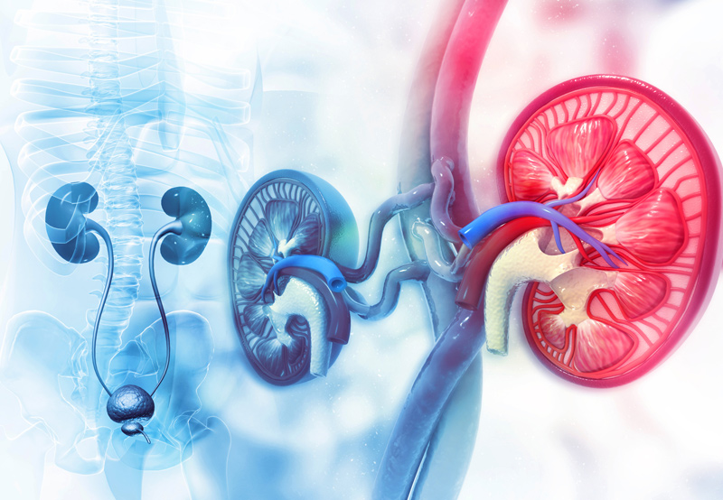

Ureteral reconstruction is done through a variety of methods, depending on the location of the injury, length of the injury, type of injury, complicating factors, and other medical conditions. A patient may need ureteral reconstruction due to radiation, pelvic surgery, endoscopic stone procedures, retroperitoneal fibrosis, and cancers of the urinary tract.
What is the Ureter?
The ureter is a long, thin tube that connects the kidney to the bladder on each side of the body. Each ureter is about eight to ten inches long and surrounded by muscle that tightens and relaxes, helping propel urine towards the bladder.

Methods Used in Ureteral Reconstruction
The six different methods used in ureteral reconstruction include:
- Cystoscopy with Ureteral Dilation and Stent Placement: This approach is for patients who aren’t good candidates for definitive repair. Surgery requires exchanging the stents due to encrustation and occlusion.
- Ureter Reimplantation: During surgery, the ureter is rerouted above the level of obstruction and repositioned into the bladder in a new location. This eliminates the need for complex bladder and bowel reconstruction. Patients with lower ureter problems may benefit from this approach. However, ureter reimplantation can’t be done for upper obstructions.
- Psoas Hitch/Boari Flap Repair: Patients with injuries above the bladder but higher in the pelvis may have extensive ureter injury and require more complex repair at the location where the bladder bridges the distance. The loss of length is accomplished by moving the upper ureter and stretching it down — along with freeing the bladder — and moving it towards the injured side. Moving the bladder will require fixing it into position by “hitching” it alongside the psoas muscle in the pelvis. This may be done in addition to a bladder flap, in which part of the bladder is tubularized to bridge the distance. Potential complication of this surgery is bladder frequency and urgency due to the bladder’s role in the repair. This is usually temporary and can be alleviated with medications.
- End-to-End Repair (Uretero-ureterostomy): A uretero-ureterostomy (UU) involves removing the diseased portion of the tube, opening up the cut ends, freeing up the length of the tube to achieve mobility, and sewing them together. During surgery, only short sections of the tube can be removed. Because of this, longer strictures and blockages cannot be repaired with this technique. Advantages of this approach include maintaining normal anatomy, avoiding complex procedures, and long-term success and patency.
- Ureter Cross Over (Trans Uretero-ureterostomy): The Trans UU is done to bypass the distal ureter by crossing the diseased ureter over the body’s midline and connecting it to the healthy contralateral side. This procedure is a great option for patients who have undergone previous complicated pelvic surgery that makes it difficult to obtain treatment with other surgical techniques.
- Ileal Ureter (Bowel Interposition): This approach to ureter reconstruction utilizes a section of the small bowel to replace longer segments of the diseased or injured ureter. This technique is reserved for patients with longer ureteral problems who have not experienced relief after more conservative approaches. Because the bowel is being used to replace a section of the urinary tract, the patient’s urine may look cloudy or contain mucus after surgery.
Robotic Pyeloplasty
This surgery is done to repair the ureter, which is the tube that attaches the kidney and bladder. Robotic pyeloplasty treats ureteropelvic junction (UPJ) obstruction.
During surgery, the surgeon makes multiple small incisions between eight and ten millimeters and inserts instruments to fix the UPJ obstruction by cutting out the narrow, scarred portion and reconnecting the normal tissue. Compared to other surgical treatments for UPJ obstruction, robotic pyeloplasty is highly effective.
